Naming of Regions of the Shoulder
We refer to findings in 3 zones around the shoulder. See image below:
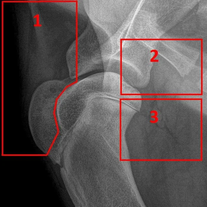
Any finding in Region #1 is termed simply a “Cranial Anomaly”. Region #2 is refered to as “Caudal Capsule” and region #3 is referred to as “Caudal Sac”. For example, the image below shows what is most probably a mineralization in the supraspinatus tendon, but we will simply call it a “Cranial Anomaly”.

Example of a cranial anomaly
Which Shoulder?
We assume that the radiograph has been taken in the standard way: the leg of interest against the table, and the opposite leg “pulled back”. Hence, the shoulder most cranial in the image is the one that will be analyzed.
A Note on our Circling of Findings
The red ellipse that we display is meant as a quick visual guide to locate a detected condition. Its exact location and shape may be approximate. It is not intended that these ellipses have a precise spatial meaning - they are just there to alert you visually that something was recognized. Also, there is only one such ellipse drawn per condition - and it is not guaranteed to encircle all occurrences of that condition, but rather, to highlight one instance. Note that with editing tools we allow you to adjust the location and shape of these ellipses (also you can completely remove them or add them).
Known Issues
The Osteochondroma finding is sometimes in error and is triggered by the end on view of the axillary artery and vein. They are surrounded by fat, so this gives them contrast.
The Fractured Supraglenoid Tubercle finding is not totally reliable as our training set does not contain enough examples, but we will be adding more data soon, and performance will improve.
The Humeral Sclerosis finding is not active yet - it will always be ‘yellow’ until our next release.